
Same MRI, Different Plan
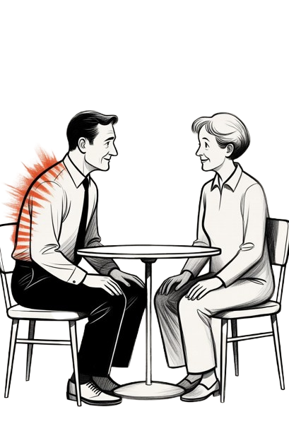
Two clients walk into the clinic in the same week, both carrying an MRI report that reads almost identically: L4-L5 disc bulge, mild facet arthropathy, some loss of disc height. One of them gets a plan built around graded MedX spinal strengthening and manual and muscular therapy. The other starts with spinal traction/decompression and Piezowave shockwave therapy before strengthening even enters the picture. Both clients have asked me some version of the same question: if our scans look the same, why doesn't our treatment?
The MRI Is Not the Diagnosis
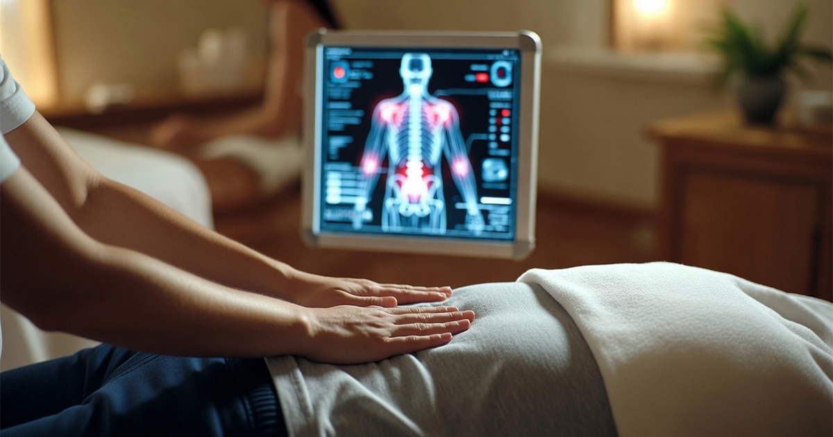
The misconception is that an MRI is a diagnosis. It is not. An MRI is a photograph of tissue at a single moment, and it says nothing about how that tissue behaves under load, how the surrounding musculature is firing, or what a person's spine actually does when they bend to load a dishwasher or twist to grab something off a truck. A disc bulge on film can belong to someone who is pain-free and fully functional, and an identical bulge can belong to someone who cannot stand for ten minutes. The scan is one data point. It is not the plan.
The Mechanics an MRI Cannot Show
Here is the mechanical piece that gets skipped in a five-minute consult: pain and dysfunction come from how forces move through the spine, not from what a static image shows. Graves 1994 found that isolated lumbar extensor strength improved by 23.5% when training included pelvic stabilization, and by -1.2%, essentially nothing, without it. Same exercise, same muscle group, different mechanical setup, completely different physiological result. If two people with identical MRIs also have different pelvic stability, different hip mobility, different years of compensatory movement patterns, they are not the same case. The film matches. The mechanics do not.
San Juan 2005 adds another layer. Multifidus activation was 51% greater with pelvic stabilization than without it. The multifidus is the small stabilizing muscle that sits close to the spine and, when it is weak or inhibited, tends to let the surrounding structures take load they were not built to carry. Two clients with the same disc finding can have very different multifidus function, one compensating well, one not. That difference alone can justify two entirely different starting points in a plan, even though the radiologist wrote the same paragraph for both of them.
What History Adds That the Scan Cannot
Then there is history, which no scan captures. A client who has been guarding a hip for six years after an old injury has built movement habits that a fresh disc bulge does not explain. A client whose job requires eight hours of sitting has a different baseline of hip flexor and psoas tension than someone who is on their feet all day, a difference explored further in why a tight psoas can keep the low back and pelvis stuck. Age, prior surgeries, activity level, sleep, stress load on the nervous system: none of it shows up on the MRI, and all of it changes what the tissue can tolerate and how fast it can adapt to loading. Treating the film instead of the client is how two people end up doing the same generic exercise sheet and getting two very different outcomes, which is part of why core workouts alone do not fix back pain for everyone who tries them.
Still living around your pain?
Let's change that. Call us and describe what you're dealing with — we'll tell you honestly if we can help.
Call 425-758-4619
Why Sequence Matters More Than the Scan
This is also why a plan has to be built in sequence, not handed over as a single prescription on day one. Someone with significant nerve involvement and high irritability may need traction/decompression and manual and muscular therapy to calm things down before their body can tolerate loaded strengthening work. Someone with a similar scan but low irritability and reasonable baseline function might go straight into supervised MedX strengthening because their tissue is already ready for load, a distinction covered in more depth in why MedX spinal strengthening works. Nelson 1995 tracked 627 completers out of 895 chronic low back pain patients and found 76% excellent or good outcomes, with 94% of those good or excellent results holding at one year. That kind of outcome does not come from a one-size-fits-all protocol matched to a scan. It comes from a plan that gets adjusted to the person doing the work, not the picture on the screen.
The Scan Opens the Conversation
None of this means imaging is useless. It rules things out, flags red flags that need a physician, and gives us a starting reference. But it was never designed to write a rehabilitation plan, and clients who expect it to are often disappointed when their identical-looking scan does not produce an identical-looking recovery. The honest answer is that the MRI opens the conversation. The client's actual mechanics, history, and current function are what close it.
You don't have to live with this. Backs and necks can get stronger, pain can ease, and the body is more capable of recovery than most people are told. This blog is here to give you honest, practical information about what actually helps. As we learn more, we'll keep sharing what works. If something here spoke to you, that's a good reason to reach out and start the first conversation.
Trent Shuford, LMT, Cert. MedX Tech.